


Abstract
Nasal administration is investigated as an effective mucosal site suitable for non-invasive vaccine delivery and able to elicit both systemic and mucosal immunity. Delivery of proteins across the epithelial barrier is a key point to induce an immune response. In this framework, nanogels are properly designed to present the delivered antigen to the immune system, promoting their targeting to specific immune populations. Nanogels for nasal administration based on dextran sulfate and ϵ-poly-L-lysine have been prepared by polyelectrolyte complexation. To unlock the potential of mucosal vaccination, we evaluated nanogel for their ability to enhance antigen delivery across mucus barriers and promote immune activation. Using fluorescein-labelled ovalbumin as a model antigen, we investigated how nanogel governs antigen transport, cellular uptake, and presentation. Produced results showed that nanogel combines favourable physicochemical properties with mucoadhesive behaviour, enabling efficient interaction with the mucus layer. This translates into enhanced uptake and controlled intracellular trafficking in murine Bone Marrow-derived-Dendritic Cells, ultimately promoting effective antigen cross-presentation. Importantly, intranasal administration in vivo revealed prolonged antigen retention within the airway mucosa compared to the free antigen, indicating sustained local antigen availability. In addition, studies in human monocyte-derived macrophages provided preliminary information on the inflammatory response and intracellular fate of the nanogels in a human-relevant system. Together, these findings contribute to the preliminary preclinical characterization of the nanogel platform, while further studies are required to establish its immunogenicity and vaccine efficacy.
Graphical Abstract

Introduction
Vaccines represent the most effective approach of preventing infectious diseases [1]. However, despite the huge advances achieved in the past years, many vaccines are still missing to fight important pathologies and to prevent emerging diseases. The current trend is to identify protective antigens and design peptide-based vaccines able to optimize antigen presentation, induce broad protective immune responses and offer a better immunogenicity/risk ratio. However, in many cases, the antigen itself is weakly immunogenic; therefore, the use of novel adjuvant or delivery systems is needed to boost the immune response [2, 3]. Delivery of proteins within the epithelial barrier is a key point to induce an immune response. In this framework, polyelectrolyte nanogels (nGEL), swollen networks composed of hydrophilic polyionic polymers, are promising delivery systems that can be properly designed to present the delivered antigen to specific immune cell populations [4]. As usual for drug delivery systems, intrinsic properties of polymeric nGEL, such as material chemistry, size, shape, surface charge, and hydrophobicity/hydrophilicity, may be determining factors in influencing the induced immune response [5]. These systems can protect vaccine antigens from degradation in vivo and induce not only strong and long-lasting antibody responses, but also potent cell-mediated immunity based on CD4 + and CD8 + T-cell responses [6].
In addition to nGEL-related properties, the route of administration critically influences the capacity of the delivery system to elicit a robust antigen-specific immune response [7]. Among the various mucosal routes, nasal administration has been extensively explored over recent decades as a promising strategy to induce both systemic and mucosal immunity [8]. Since the early 2000s, following the introduction of the first commercially available intranasal influenza vaccine, FluMist (approved in the United States in 2003), the nasal route has garnered increasing interest as a viable alternative to conventional parenteral vaccination. In this context, the nasal cavity represents a highly attractive target for vaccine delivery, as it constitutes a primary entry site for numerous pathogens while simultaneously offering a non-invasive and patient-friendly approach, thereby enabling needle-free immunization and facilitating mass immunization campaigns [8].
Mucosal vaccination has emerged as an attractive strategy against pathogens that primarily invade the host through respiratory, gastrointestinal, or genitourinary mucosal surfaces [9,10,11,12,13,14]. Unlike conventional parenteral vaccines, which predominantly induce systemic IgG responses, mucosal immunization can elicit both systemic and local immune responses directly at the site of pathogen entry [9, 10, 13, 14]. This is particularly relevant for respiratory pathogens, where the induction of local immunity may provide additional protection by preventing pathogen attachment, colonization, and early replication at the mucosal surface, responses that are less efficiently induced by parenteral vaccination. The ability of mucosal vaccines to induce IgA, is considered a key advantage, as IgA can neutralize pathogens before they cross the epithelial barrier and establish infection [9, 13, 14]. Furthermore, mucosal vaccination promotes the generation of tissue-resident memory B and T cells within the respiratory tract, enabling rapid and long-lasting immune responses upon pathogen re-exposure [9, 10]. Collectively, these features may contribute not only to protection against disease but also to a reduction in pathogen transmission by limiting infection at its portal of entry [9, 12].
Based on these considerations, in our previous study, polyelectrolyte nGEL based on ϵ-poly-L-lysine (ϵPL) and dextran sulfate (DS) were conceived by a design of experiment (DoE) approach for ovalbumin (OVA) delivery as antigen carrier for nasal vaccine therapy [4]. The green preparation method yielded stable nGELs with particle mean size (Z-Ave) ~ 150 nm, polydispersity (PDI) ~ 0.2, and zeta potential (ZP) ~ + 40 mV), which maintained their physicochemical properties for up to 6 months at 4 °C. The optimized nGEL exhibited no cytotoxicity in HFF1, NCTC, and Calu‑3 cells at ≤ 0.117 mg/mL, supporting its suitability for nasal delivery. Moreover, OVA was efficiently encapsulated (EE 95%) after 24 h of incubation time, and the resulting OVA_nGEL retained physicochemical properties appropriate for nasal administration [4]. Therefore, in order to deeply explore and evaluate the applicability of nGEL for mucosal antigen delivery, in this work we aimed to encapsulate fluorescein isothiocyanate-ovalbumin (OVA-FITC) in the nGEL to in vitro investigate uptake kinetics and intracellular trafficking of nGEL for antigen presentation in murine Bone Marrow-derived-Dendritic Cells (BMDCs) and to track its distribution in vivo.
The formulation was characterized by a physico-chemical and technological point of view in order to determine the Z-Ave, PDI and ZP. In addition, the mucoadhesive behaviour and strength of nGEL was evaluated in vitro to demonstrate the adhesion-mediated penetration in the mucus. Uptake kinetics and intracellular trafficking of nGEL were investigated by confocal microscopy in BMDCs, and OVA257−264 specific T-Cell hybridoma was used to assess the antigen cross-presentation efficacy. Moreover OVA-FITC_nGEL were administered in mice by intranasal instillation to track the residence time of the antigen in airway mucosa with respect to its free form in vivo. Finally, in vitro activation by nGEL and their subsequent phagocytosis in human monocyte-derived macrophages (MDMs) were assessed to provide a preliminary evaluation of their biocompatibility in a human cell model.
Materials and methods
Materials
Epsiliseen®-H (ɛ-poly-L-lysine, ɛPL, MW 4.7 kDa) was kindly gifted by Siveele B.V. (Breda, Netherlands). Dextran sulfate sodium salt (DS, Mr ~ 40.000), hydrogen chloride (HCl, reagent grade, 37%), sodium hydroxide (NaOH), salts, albumin from chicken egg white (OVA) and mucin from porcine stomach type II were purchased from Merck (Darmstadt, Germany). Ovalbumin fluorescein conjugate (OVA-FITC) was purchased from Invitrogen (Thermo Fisher Scientific, Waltham, MA, USA).
Preparation of optimized DS-ϵPL nGEL
DS solution in water at specific concentration (1 mg/mL) and ϵPL solution in water (1.25 mg/mL) were prepared followed by pH adjustment by addition of 1 N HCl or 0.05 N NaOH solution. DS aqueous solution was added dropwise to ϵPL aqueous solution (volume ratio 1:3) at room temperature under magnetic stirring (55 xg). nGEL were formed instantaneously, as confirmed by the increase of the turbidity of the mixture. The suspension was stirred for 1 h and then nGEL were separated by centrifugation at 16,128 xg for 30 min at 4 °C (ThermoScientific™ SL16R, ThermoFisher Scientific, Waltham, MA, USA). Finally, the pellet was resuspended in distilled water and used for further studies.
Preparation of OVA-FITC loaded DS-ϵPL nGEL
OVA-FITC_nGEL was prepared following the above method. OVA-FITC (0.3 mg/mL) was firstly mixed with the polycation, and then the negatively charged DS solution was added dropwise to form a stable polyelectrolyte nGEL (OVA-FITC_nGEL). The complex was incubated under magnetic stirring (55 xg) at 25 °C for 24 h to allow the complete OVA-FITC loading into the nGEL network. The formulation was independently prepared in three separate batches and results were expressed as the mean ± standard deviation (SD).
OVA-FITC encapsulation efficiency
In order to determine the protein encapsulation efficiency (EE%), the obtained OVA-FITC_nGEL was centrifuged at 12,000 for 30 min at 4 °C, and the amount of unbound OVA-FITC in the supernatant was determined using BCA Protein Assay Kit (Merck, Milan, Italy). The protein standard curve was obtained following the microassay indicated by the manufacturer. The calibration curve for the quantitative evaluation of OVA-FITC was linear in the range 6–1000 µg/mL (R2 = 0.9964). Supernatant from centrifugation of an empty nGEL was used as a negative control. The results were expressed as the mean ± standard deviation (SD) of three independent batches.
Particle size, polydispersity index, and zeta potential measurements
Mean particle size (Z-Ave) and polydispersity (PDI) were determined by dynamic light scattering (DLS), whereas zeta potential (ZP) measurements were performed by electrophoretic light scattering (ELS) using a Zetasizer Nano S90 (Malvern Instruments, Malvern, UK) at a detection angle of 90°, at 25 °C with a 4 mW He–Ne laser operating at 633 nm.
Each analysis was performed in triplicate on three independently prepared batches and results were expressed as the mean ± standard deviation (SD).
Mucoadhesive evaluation of optimized DS-ϵPL nGEL
In vitro methods were used to assess the mucoadhesive properties of the optimized nGEL. Briefly, mucin (0.1% w/v) was suspended in simulated nasal fluid (SNF: 2.192 g NaCl, 0.145 g CaCl2 and 0.745 g KCl in 250 mL of double distilled water; pH 5) and stirred overnight to allow its complete dispersion. The interaction between nGEL and mucin was determined by mixing equal volumes of mucin dispersion and sample suspension for 15 min at 25 °C. After 1 and 24 h of incubation at 37 °C, turbidimetry and mucin particles method were performed. In particular, turbidimetric measurements were evaluated comparing the absorbances at 650 nm by UV-VIS spectrophotometer (UH5300 UV-Visible Double-Beam Spectrophotometer, Hitachi Europe, Milan, Italy) of the native mucin and each dispersion. For the nGEL-mucin interaction study, variation in Z-Ave, PDI and ZP were considered after incubation of nGEL with mucin, using a Zetasizer Nano S90 (Malvern Instruments, Malvern, UK). Each analysis was performed in triplicate on three independently prepared batches and results were expressed as the mean ± standard deviation (SD).
Mucoadhesive strength by binding efficiency
nGELs were evaluated regarding their mucoadhesive strength based on the interaction with the negatively charged mucin. Briefly, equal volumes of mucin (0.1% w/V in SNF) and nGEL nanosuspension were stirred for 15 min at 25 °C and incubated for 1 and 24 h at 37 °C, and then centrifuged at 16,128 xg for 1 h at 25 °C. UV-VIS spectrophotometer (UH5300 UV-Visible Double-Beam Spectrophotometer, Hitachi Europe, Milan, Italy) was used to quantify the amount of free mucin in the supernatant at 260 nm. The calibration curve for the quantitative evaluation of mucin was linear in the range 93.55–1000 µg/mL (R2 = 0.998). The mucin-binding efficiency (BE%), expressing the mucoadhesive strength of the nGEL, was calculated according to Eq. (1):
Each analysis was performed in triplicate on three independently prepared batches and results were expressed as the mean ± standard deviation (SD).
In vitro biological evaluation
Human monocytes isolation and macrophages differentiation
Peripheral Blood Mononuclear Cells (PBMC) were isolated from leukocyte-platelet concentrates (buffy coat) obtained from healthy donors after appropriate informed consent. Cell separation took place by density gradient generated by Ficoll-Paque (GE Healthcare, Bio-Sciences AB, Uppsala, Sweden), and monocytes isolation was obtained by positive selection for CD14 with magnetic microbeads (Miltenyi Biotec, Bergisch Gladbach, Germany). Isolated monocytes were seeded at a density of 5 * 105 cell/mL in 24-well flat bottom plates (Corning® Costar®; Corning Inc. Life Sciences, Oneonta, NY, USA), and cultured in RPMI 1640 + Glutamax-I medium (GIBCO by Life Technologies, Paisley, UK) enriched with 5% heat inactivated human AB serum (Saint Louis, MO, USA), 50 µg/mL gentamicin sulfate (GIBCO) (hereafter referred to as the complete medium) at 37 °C in a humidified incubator containing 5% CO2. To generate human monocyte-derived macrophages (MDMs), monocytes were exposed for 7 days at 50 ng/mL human Macrophage Colony-Stimulating Factor (M-CSF, R&D Systems, Minneapolis, MN, USA) in the complete medium, and medium changes were performed every 3 days.
Bone marrow-derived-dendritic cells (BMDCs) generation
C57BL/6 (eight-week-old, female) were purchased from Charles River (Lecco, Italy) and housed at IGB-CNR Animal House Facility under standard pathogen-free conditions abiding institutional guidelines. The experiments were performed in accordance with the EU Directive 2010/63/EU for animal experiments, and where authorized by Italian Ministry of health with the authorization number 7E58D.12 released on May 20, 2020. Precursors for BMDCs derivation were isolated from the tibiae of euthanized C57BL/6 mice. Tibiae ends were cut, and bone marrow was flushed with the needle of a syringe filled with ice-cold RPMI 1640 medium (Microgem srl, Naples, Italy). Cells were washed twice with medium and clusters of cells were disaggregated by pipetting. BMDCs precursors were plated in 10 mm untreated plates with 200 U/mL recombinant murine granulocyte/macrophage colony-stimulating factor (GM-CSF, Peprotech, NJ, USA) in RPMI 1640 medium supplemented with 10% foetal calf serum (FCS), 60 µg/mL penicillin, 100 µg/mL streptomycin, 1 mM sodium pyruvate and 50 µM 2-mercaptoethanol (GIBCO by Life Technologies, Paisley, UK). GM-CSF was added on day 1–3 and 6 after plating, replacing the culture medium. Immature DCs were collected eight days later and used in antigen presentation assays. For confocal microscopy experiments, BMDCs precursors were plated on sterile glass coverslips of 12 mm diameter (Waldemar Knittel, Germany) in 24-well cell culture plate on day 1 of culturing.
Human primary monocytes and macrophages activation
Before addition to cell culture, nGELs were pre-incubated in 50% inactivated human AB serum at 37 °C for 1 h in a continually shaken, in order to obtain the formation of a bio-corona of serum proteins and other components on the particle surface thereby ensuring particle stability in culture. The serum-nGEL mixture was added directly to culture wells, adjusting nanoparticle and serum concentration to the desired values for each treatment. For dose-response experiments, monocytes and MDMs, isolated from 2 different donors, were exposed to culture medium alone (medium/ negative control), or to 5 ng/mL lipopolysaccharide (LPS) (positive control; from E. coli O55:B5; Sigma-Aldrich, Saint Louis, MO, USA), or to serum pre-coated nGEL (concentration range 0.2–5 µg/mL). After 24 h of incubation at standard cell culture condition, culture supernatants were collected and stored at -80 °C for cytokine/chemokine analysis by ELISA (R&D Systems, Minneapolis, MN, USA).
Antigen presentation by murine dendritic cells
BMDCs (5 * 105/well in 0.1 mL) were incubated in a 96 well cell culture plate with described concentrations of nGEL, OVA-FITC loaded_nGEL or free OVA-FITC. As a control, BMDCs incubated only with medium were used. After overnight incubation, 5 * 105 /well in 0.1 mL B3Z cells (OTI hybridoma line) were added for co-culture. B3Z is an OTI T hybridoma cell line, able to recognize the OVA257–264 SIINFEKL peptide. B3Z cell culture medium is composed by RPMI1640 and 10% FCS, 100 U/mL penicillin, 100 µg/mL streptomycin 1% glutamine, 1% non-essential amino-acids (GIBCO by Life Technologies, Paisley, UK), 1% sodium pyruvate, 50 µM 2-mercaptoethanol. After 40 h co-culture, cell culture supernatant (0.1 mL/well) was collected and used for IL-2 detection by ELISA assay using the ELISA MAX™ Deluxe Set Murine IL-2 (BioLegend, Canada, US) according to the manufacturer’s instructions. Supernatants of co-cultures were assayed in duplicate; results are representative of three independent experiments.
Uptake kinetics and internalization of NG in human macrophages and murine BMDCs
MDMs or BMDCs, plated on glass coverslips at the density of 3 × 10⁵cell, were treated with OVA-FITC_nGEL (25 µg/mL) or free OVA-FITC (25 µg/mL) at different time points (30 min; 2 h; 6 h). At the end of the incubation, cells were fixed with 4% paraformaldehyde for 15 min, washed three times in PBS and then incubated in blocking solution [0.5% (w/V) BSA, 50 mM NH4Cl in PBS, pH 7.4, 0.05% saponin] for 30 min at 25 °C. Cells were subsequently incubated with the specified antibodies diluted in blocking solution O/N at 4 °C. After incubation with the primary antibody, cells were washed three times in PBS and incubated with the respective secondary antibody diluted in blocking solution (AlexaFluor-488 or -568 conjugated, Thermo Fisher Scientific Inc., Invitrogen, Bremen, Germany). Nuclei were stained with Hoechst 33,258. Finally, cells were washed 3 times in PBS and once in sterile water to remove salts. Coverslips were then mounted on glass-microscope slides with Mowiol (20 mg mowiol dissolved in 80 mL PBS). Images were taken using a Zeiss-LSM 700 confocal microscope equipped with a 63x/1.4 NA oil-immersion objective. Optical confocal sections were taken setting a pinhole to 1 Air Unit.
The intracellular fate and localization of the nanoparticles were investigated using the following antibodies: Anti-LAMP-1 (PA1654A, Thermo Fisher Scientific Inc.; 1:400) lysosomal marker; Anti- EEA1 (# 14-9114-82, Thermo Fisher Scientific Inc.; 1:100), early endosome marker; Anti-Rab11 (D4F5) XP ( # 5589 S; Cell Signaling Technology’s, Danvers, MA, USA; 1:100), recycling endosome marker; anti-Rab7 (#R4779, Sigma-Aldrich, Saint Louis, MO, USA; 1:100), late endosome marker; anti-Calnexin (C5C9) (#2679; Cell Signaling Technology’s, 1:100), ER marker; anti-LC3 A/B (#12741; Cell Signaling Technology’s, 1:100), autophagosome marker.
In vivo studies
Animals
Animal experiments were carried out following the EU Directive 2010/63/EU for animal experiment guidelines and in agreement with the principles and procedures outlined by the Bio Ethical Committee of Quaid i Azam University (approval: BEC-FBS-QAU 2026/404).
C57BL/6 male mice aged 8 weeks (24 ± 0.35 g) were purchased from the National Institute of Health (NIH), Islamabad, Pakistan and they were maintained at standard conditions of both temperature (20 ± 2 °C) and humidity (65%) following a 12 h light/12 h dark cycle (light on 8:00 a.m.) with food and water ad libitum.
Real time fluorescence optical imaging
Experimental groups were designated as follows: free OVA-FITC (n = 5), OVA-FITC_nGEL (n = 5), unloaded nGEL (n = 5) and PBS as control (n = 5). Mice were administered with 30 µL by nasal instillation, 15 µL in each nostril.
The real time fluorescence optical imaging was carried out through Bruker In-Vivo Xtreme X-ray/optical imaging system in order to evaluate the interaction and the mean residence time of the various formulations in the nasal tissue [15]. The images were acquired before the administration, immediately after administration (T0) and at 0.5, 1, 2, 6 h following nasal administration. Acquisitions and analyses of images were performed by the Bruker MI software (Bruker, Billerica, MA).
Isolation of freshly nasal tissue
Mice were euthanized by inhalation of 5% isoflurane in oxygen (at a flow rate of 1 L/min) until respiratory arrest occurred, followed by cervical dislocation. Isolation of freshly intact mouse nasal tissue with preserved anatomical organization was performed as previously reported [16].
Briefly, after separation of the entire head from the rest of the body, the skin and fur were removed from the head exposing the skull. From here on, the entire procedure was performed by keeping the sample on a piece of Kimwipes, soaked in cold PBS and placing it on ice. Occasionally, the sample was moistened with cold PBS solution to maintain cell and tissue viability. Under a dissection microscope, the skull bone covering the brain and olfactory bulbs, was removed by making a small incision in the occipital area of the skull, gently lifting and trimming off the skull bone. The right and left nasal bones were removed by making an incision in the central region of the frontal bones, just caudal to the nasal bones, and cutting the bone laterally to facilitate removal. The anterior portion of the right and left zygomatic arches were broken using the rongeur, and the medial suture between the right and left nasal bones was loosened with a razor blade. Thus, the nasal bone was gently removed by grasping its caudal end with serrated forceps separating it side to side from the underlying tissue. Once separated from the underlying tissue, the bone was cut with scissors at the rostral end. Afterward, the sample was positioned ventrally to identify and break, with a serrated forceps and a rongeur, the vomer bone, the anterior portion of both incisors and the articular region of the right and left anterior maxilla. Thus, the nasal tissue underneath the jaw was gently separated and the bone fragment lifted. Finally, the sample was repositioned dorsally, and a stab incision was made to separate the entire olfactory epithelium and nasal tissue from the olfactory bulbs and brain. Samples were stored in cold PBS or 4% formaldehyde for subsequent analyses.
Statistical analysis
GraphPad PRISM 10.1.0 (GraphPad Software, Inc., La Jolla, CA, USA) was used for statistical analysis of data produced; significance was calculated applying p < 0.05 as the minimum level. For mucoadhesive studies, two-way ANOVA was performed; multiple comparisons were performed according to Tukey’s multiple comparisons test for the turbidimetric assay, and for Z-Ave, PDI and ZP data.
For ELISA assay, two-way ANOVA followed by Dunnett’s multiple comparison test was performed.
For real time fluorescence optical imaging, all results were expressed as mean ± SD. Normality was tested using Shapiro–Wilk normality test. For in vivo studies, normally distributed data were analysed by two-way repeated measures ANOVA with the Geisser-Greenhouse correction, followed by Tukey’s test.
Results and discussion
Physico-chemical characterization of OVA-FITC_nGEL
Recently, nanocarriers prepared by ionic crosslinking of polyelectrolytes have been studied in a wide range of biomedical applications (i.e., colonic drug targeting, protein or lipophilic compounds delivery, theranostic) [17,18,19,20]. Polyelectrolyte complexation represents a sustainable strategy for synthesizing nGEL, achieved through ion pairing between polyanions and polycations in aqueous media [21]. The electrostatic interactions of oppositely charged polyions are increasingly recognized as a promising route for the development of nGEL systems [22]. The obtained system possesses high colloidal stability, while its size and charge can be optimized for the specific purpose [4].
Here, we propose a simple nGEL obtained by polyelectrolyte complexation, based on two polyions derived from a naturally occurring highly biocompatible polysaccharide, DS, and a cationic homopolymer of L-lysine, ϵPL, naturally produced by Streptomyces albulus, which instantly and spontaneously self-assembles to form nGEL particles [23, 24].
The nGEL is formed through electrostatic complexation between the negatively charged sulfate groups (–OSO3−) of DS and the positively charged protonated ε-amino groups (–NH3+) of ϵPL, leading to spontaneous self-assembly and stabilization of the nGEL structure. Prior to mixing, the pH of the individual DS and ϵPL aqueous solutions was adjusted to modulate the ionization of the interacting groups.
In order to evaluate the in vitro intracellular trafficking of nGEL and the in vivo biodistribution after nasal administration in mice, the optimized nGEL was loaded with the fluorescent form of OVA [4]. The fluorescent protein OVA-FITC was loaded into nGEL (0.3 mg/mL) by polyelectrolyte complexation method: OVA-FITC was added to the polycation phase prior to polyanion dripping, since the pH of the environment (pH ~ 5) is close to the protein pI (4.9), thus the net charge of the molecule was neutral, consequently avoiding any interaction with ϵPL. This condition was selected to minimize competitive electrostatic interactions between OVA-FITC and the positively charged ε-amino groups of ϵPL, thereby preserving the availability of the polycation for complexation with the negatively charged sulfate groups of DS. This step was followed by the polyanion addition into the polycation solution leading to the occurrence of electrostatic interactions between the two polyions that held the protein within the polymeric network. The final pH of the resulting nGEL dispersion was 5.42 ± 0.03. Therefore, OVA-FITC is expected to carry a slight net negative charge in the final formulation and its incorporation is likely governed by a combination of physical entrapment within the rapidly forming DS/ϵPL network and residual electrostatic and non-covalent interactions contributing to antigen retention within the nanogel matrix.
Homogeneous formulation was obtained (PDI < 0.3), with a Z-Ave of 204.1 ± 0.5 nm, slightly higher than the value observed for the blank system (Z-Ave = 155.2 ± 2.9 nm; PDI < 0.2) due to the presence of the fluorescent molecule. The effective incorporation of OVA-FITC into the nGEL network was assessed by BCA assay resulting equal to 81.65% ± 0.20, thus corroborating previous results obtained for OVA-nGEL [4]. The protein was also partially adsorbed into the nGEL surface as revealed by the reduction of ZP from + 45.10 ± 2.30 mV for the empty system to + 31.9 ± 0.9 mV after the addition of OVA-FITC. Although protein loading resulted in a reduction of the surface charge, the ZP remained higher than + 30 mV, suggesting that sufficient electrostatic repulsion was maintained to prevent particle aggregation. These features further support the suitability of the developed nGEL as antigen carriers for intranasal vaccine delivery. Indeed, nGEL obtained by polyelectrolyte complexation offer several advantages, including preparation under mild aqueous conditions, avoidance of organic solvents, and preservation of protein integrity during formulation. In addition, their hydrated polymeric network can efficiently accommodate biomacromolecules while maintaining favorable physicochemical properties, as previously demonstrated [4]. The nanometric size of the particles is expected to facilitate interactions with the nasal mucosa and antigen-presenting cells, whereas the low PDI indicates the formation of a homogeneous and reproducible nanosystem. Moreover, the highly positive surface charge not only contributes to colloidal stability but may also enhance electrostatic interactions with the negatively charged mucosal environment, potentially increasing residence time at the administration site. Overall, these characteristics are particularly attractive for mucosal vaccine delivery, where prolonged antigen retention and efficient uptake by immune cells are key prerequisites for the induction of an effective immune response. Considering the intended mucosal application, the mucoadhesive property of nGEL suspension was evaluated prior to investigate the in vitro and in vivo behaviour as candidate carrier for vaccine therapy.
Evaluation of nGEL-mucin interaction and complex stability
A major drawback of nasal drug delivery is the rapid mucociliary clearance, which primarily serves to protect the respiratory tract from harmful inhaled substances. However, this mechanism also restricts drug absorption, as conventional formulations exhibit a clearance half-life of approximately 15 min. Consequently, this significantly reduces or even eliminates therapeutic efficacy [25]. The nasal epithelial mucus consists of roughly 90–95% water, 2–5% mucins, about 1% salts, and varying amounts of proteins and lipids [26]. Among these components, mucins primarily determine the physicochemical properties of mucus. These mucins are highly glycosylated proteins featuring a linear peptide backbone with oligosaccharide side chains. Terminal sialic acid and sulfate groups on these chains confer a negative charge to the glycoprotein [27].
Mucoadhesion, which enables prolonged retention, is often regarded as a key requirement for effective nasal drug delivery. Incorporating mucoadhesive polymers into nasal formulations can enhance their residence time within the nasal cavity improving contact with the absorptive mucosal surfaces.
Based on these considerations and taking into account the intended mucosal application, the mucoadhesive and interactive properties of nGEL were assessed using three complementary approaches: the nGEL-mucin interaction study, turbidimetric analysis, and binding efficiency evaluation. To evaluate the interaction between nGEL and mucosal components, a mucin interaction study was performed. Although physiological nasal mucociliary clearance is a rapid process typically occurring within 15–20 min, the evaluation of nGEL–mucin interactions was extended to 1 h and 24 h not to simulate in vivo mucosal residence time, but rather to assess the colloidal stability and structural integrity of the formed complexes over time [28] (Fig. 1).

Mean particle size (Z-Ave) (A), PDI (B) and Zeta potential (ZP) (C) values of nGEL after 1 and 24 h of incubation with mucin (nGEL-MUC) at 37 °C. Significance was set at ****p ≤ 0.0001
The obtained results showed the presence of interactions between nGEL and the glycoprotein, as evidenced by the significant increase in particle size and the variation of ZP of the nGEL incubated with mucin (nGEL-MUC) at the considered time points. Interestingly, while pure mucin exhibited a higher apparent hydrodynamic size (905.3 nm ± 29.27) than the complex, this behavior can be ascribed to the intrinsic tendency of free mucin macromolecules to form large, loose, and open supramolecular aggregates in aqueous dispersion, which heavily dominate intensity-based DLS measurements. Upon incubation with the nGEL, the interaction leads to a conformational rearrangement and compaction of the mucin chains, reflecting the formation of a more compact, organized, and uniform nanoconstruct rather than the swollen aggregates typical of free mucin.
This controlled interaction and structural homogeneity are further confirmed by PDI values ≤ 0.3 at all time-points (Fig. 1B), demonstrating that the system avoids chaotic precipitation or uncontrolled aggregation. Complex formation and interaction were also confirmed by turbidimetric measurements (Fig. 2).
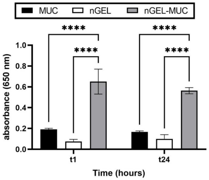
In vitro assessment of nGEL/mucin interactions after 1 and 24 h of incubation at 37 °C by turbidimetric assay at 650 nm. Significance was set at ****p ≤ 0.0001
To exclude any contribution of the nGEL’s intrinsic turbidity to the measured absorbance, the absorbance of the nGEL alone was also evaluated and included as a control (Fig. 2). The nGEL dispersion showed negligible absorbance under the experimental conditions, indicating that the increase observed following incubation with mucin was mainly attributable to nGEL-mucin interactions.
It has been reported in literature that absorbance measurement provides a rough estimation of the particle-mucin interaction [29, 30]; in case of interaction, the adsorption of mucin around the surface of the particles induces an aggregation phenomenon that can be detected as an increase in UV absorbance compared to that of native mucin [30]. The interactions at the mucin–particle interface are complex and governed by multiple mechanisms described by theories such as wetting, electronic, adsorption, fracture, mechanical, and diffusion interlocking [31]. Mucoadhesive materials are typically hydrophilic macromolecules rich in functional groups capable of forming hydrogen bonds. In this context, the observed interaction likely resulted from electrostatic forces, such as hydrogen bonding and van der Waals interactions, between the negatively charged mucin and the positively charged nGEL, primarily through adsorption. Mucoadhesion generally occurs in two stages: an initial contact (wetting) phase, followed by a consolidation phase during which adhesive interactions are established. Adsorption plays a critical role when the formulation cannot be applied directly to the target mucosa, whereas consolidation becomes essential under conditions of significant mechanical stress that could dislodge the formulation [32]. To further assess the affinity between nGEL and mucin, the binding efficiency (B.E.%) was measured at the same time intervals. After 1 h of incubation at 37 °C, the B.E.% reached 67.6 ± 0.4%, while after 24 h at the same temperature it remained stable at 55.2 ± 1.1%, confirming the robust and stable interaction between the components over time. These findings demonstrate the strong affinity of nGEL for mucosal glycoproteins, supporting its capability to interact effectively with the nasal mucosa upon administration.
Antigen processing and presentation
In order to measure the ability of nGEL to improve the antigen engulfment by antigen presenting cells and the antigen delivery to the intracellular route of processing and presentation in the MHC-I (cross-presentation), BMDCs were incubated with OVA FITC, OVA-FITC_nGEL, or unloaded nGEL and then co-cultured with OVA specific T B3Z cells. Recognition of the peptide OVA257–264 (SIINFEKL), derived from OVA processing and presented in the major histocompatibility complex (MHC-I) H2Kb by B3Z T cell receptor leads to the transcriptional activation of the IL-2 promoter elements, resulting in IL-2 production correlated with the OVA uptake and processing [33]. Results showed in Fig. 3 demonstrate that when encapsulated in nGEL, OVA-FITC is taken up by BMDCs and processed leading to OVA257−264 peptide presentation at greater extent compared to the free protein.

IL-2 production after B3Z cell activation by antigen-loaded BMDC. Different concentrations (5, 10 and 25 µg/mL) of free OVA-FITC or OVA-FITC_nGEL were used. Unloaded nGEL (5, 10 and 25 µg/mL) represents the negative control. Each value represents the average value (± SD) of three experimental replicates. Significance was set at ****p < 0.0001
Comparable results were observed with lipid-OVA nanoaggregates, which significantly enhanced IL-2 production and secretion by T cells, following OVA processing by BMDC cells [34]. Conversely, Aroffu et al. reported no detectable in vitro immune response upon incubation of antigen-presenting cells with either soluble OVA or OVA-encapsulated within transfersome [35]. Antigen cross-presentation is a crucial intracellular process carried out only by professional antigen presenting cells (APC) and corresponds to the loading of MHCI molecules by peptides derived by extracellular source and normally addressed to MHCII molecules. Exogenous proteins, after engulfment, are processed by APC and antigenic peptides are loaded on MHCI molecules prompting naïve T CD8 + cell activation, allowing them to recognize the same antigen when presented by cells infected by intracellular pathogens, leading to CD8 mediated killing [36]. Antigens can be delivered into APCs by macropinocytosis, endocytosis or phagocytosis and the intracellular trafficking significantly influences the cross-presentation efficacy: targeting to selected subcellular compartments could help the adaptive immune response activation [37]. Herein, we analysed cellular uptake and intracellular localization of OVA-FITC_nGEL and free OVA-FITC in BMDCs by colocalization with fluorescently labelled markers of different intracellular compartments. In Fig. 3 is reported the OVA-FITC internalization and localization within the cell after 6 h.

OVA-Antigen internalization and localization in murine BMDCs. Confocal images of free OVA-FITC (A-D) or OVA-FITC_nGEL (E-H) uptake (25 µg/ml) by BMDCs upon incubation for 6 h. Early endosomes are visualized with EEA1 (A and E); lysosomes with LAMP1 (B and F), autophagosomes with LC3 (C and G) or Calnexin (D and H) in red. Cell nuclei are stained blue with Hoechst. Bar=10 μm. Objective: 63x/1.4 NA oil-immersion objective. Representative images from one of three independent experiments are shown
As shown, OVA-FITC_nGEL after 6 h (Fig. 3, E-H) was internalized more efficiently when compared to the control free OVA-FITC (Fig. 3, A-D), and this is already observed after 30 min (Fig. S1, Supporting information). Once inside the cell, the intracellular fate of the two samples was quite different: OVA-FITC_nGEL displays partial colocalization with the endosomal marker EEA1 (Fig. 3E), the autophagosome marker LC3 (Fig. 3G), and the Endoplasmic Reticulum (ER) marker Calnexin (Fig. 3H), while no localization was observed in the lysosomal compartment as revealed by the marker LAMP-1 (Fig. 3F). On the contrary, free OVA-FITC was not localized in the same intracellular compartments, displaying partial colocalization only with the EEA1 (Fig. 3A) and LAMP-1 (Fig. 3B) compartments. From the reported observations, we hypothesize that free OVA-FITC, once internalized, is degraded in the lysosomal pathway and does not enter the cross-presentation pathway. Interestingly, OVA-FITC_nGEL, after internalization, was recruited to early endosomes and phagosomes and then transported to ER where it can be loaded onto MHC class I, following the pathway described by Embgenbroich and Burgdorf [38]. The ability of nGEL in protecting the loaded protein by lysosomal degradation showed in our study is corroborated by literature findings, being the antigen presentation to the MHC class I pathway higher when OVA was loaded into self-assembly nanogel prepared with cholesterol-bearing cluster dextrin, compared to the free protein [39].
Similar observations were reported by Brusco et al., who developed lipid/poly(lactic-co-glycolic acid) (PLGA) hybrid nanoparticles (hNPs) for the co-delivery of OVA and a synthetic adjuvant to the respiratory mucosa. Confocal microscopy analysis revealed that hNPs engineered with a polyethyleneimine (PEI) coating were capable of entering the endosomal-lysosomal pathway, as indicated by a peak in colocalization with the lysosomal marker LAMP-1 at 2 h post-incubation. Subsequently, a reduction in colocalization signal at 24 h suggested that the nanoparticles had escaped into the cytoplasm, likely avoiding lysosomal degradation. This lysosomal escape is considered a critical step in facilitating antigen cross-presentation [29]. It has been demonstrated that intra-endosomal antigen stability is a crucial characteristic for improving cross-presentation, and that rapid lysosomal degradation of internalized antigens negatively affects this process [34, 40].
Evaluation of the inflammatory response in human innate immune cells
In order to investigate the capability of nGEL to trigger an inflammatory immune response, human monocytes and MDMs were stimulated with unloaded and OVA-FITC_nGEL at different concentrations (0.2–5 µg/mL) for 24 h. Monocytes and MDMs are innate immune cells that participate in rapid defensive responses, primarily through the initiation and amplification of inflammatory reactions. Prototypical effectors of the inflammatory reaction have been measured, such as the inflammatory cytokines TNF-α and IL-6 and the anti-inflammatory cytokines IL-10. As shown in Fig. 4, unloaded nGEL did not significantly affect TNF-α and IL-6 production both in monocytes (Fig. 4A) and MDMs (Fig. 4B), conversely to LPS, which is a potent activator of innate immune system. Furthermore, no effect has been observed for IL-10 production in both cell types. Thus, nGEL as itself is unable to induce an inflammatory response even at high concentrations; this result is in agreement with literature studies on cholesterol-modified hyaluronic acid nanogel-based hydrogel [41]. The treatment with OVA-FITC_nGEL showed similar results, with a not significant tendency to increase cytokines production only at the highest concentration (5 µg/mL) in monocytes.

Cytokine production by human monocytes (A) and MDMs (B) cultured for 24 h in culture medium alone (medium) or containing serum-precoated nGEL (nGEL), serum-precoated OVA-FITC_nGEL and LPS. The production of TNFa, IL-6 and IL-10 was measured in the 24 h supernatants by ELISA. Data are presented as mean of three independent experiments ± SD
A nanoparticle-based system loaded with an antigen for mucosal vaccine therapy has the potential to elicit an inflammatory immune response in human monocytes and MDMs; however, the immunological profile induced by such a system depends on multiple factors, including the nanoparticle design, the nature of the antigen, the presence of adjuvants, and the intended immunological outcome [42]. Monocytes and MDMs are pivotal components of the innate immune system. If on the one hand, a controlled inflammatory activation can be beneficial for priming adaptive immunity, particularly when the vaccine aims to induce a robust T-cell-mediated response; on the other hand, excessive inflammatory activation may lead to local or systemic adverse effects [43]. For mucosal vaccines, especially those administered via nasal or oral routes, it is critical to avoid inflammation that could compromise mucosal tissue integrity [11].
Our findings corroborate those reported by Schlich et al., who demonstrated that the exposure of human monocytes to lipid-OVA nanoaggregates did not elicit any significant change in the production of the pro-inflammatory cytokines TNF-α and IL-6 [34].
This observed lack of responsiveness in human monocytes upon exposure to the nanoaggregates may be attributed to two potential mechanisms: either the cells do not detect the aggregates, or the aggregates are detected but not recognized as a danger signal, thereby failing to induce an innate immune reaction [34]. Similar findings were also found by Aroffu et al., which developed transfersomes enriched with glycerol and/or hyaluronic acid to favor skin deposition of ovalbumin [35]. The authors found no inflammatory response of OVA in solution or encapsulated in enriched transfersomes, confirming the safety of these carriers for a vaccine formulation [35]. Our nanoplatform composed by DS and ϵPL is unlikely to elicit a significant inflammatory response because the electrostatic interaction between these oppositely charged polymers forms a stable nanogel with low intrinsic immunogenicity. The biocompatibility of both DS and ϵPL, coupled with their use in forming stable nanogel, suggests a favorable interaction with immune cells. The absence of an inflammatory immune response may be attributed to several factors such as the nature of the polymers which are well-established biomaterials with demonstrated biocompatibility and safety profiles [44,45,46]. Moreover, neither DS nor ϵPL contains strong molecular motifs that would typically trigger an acute inflammatory response [46]. The absence of a marked inflammatory response in monocytes and monocyte-derived macrophages upon exposure to antigen-loaded nanogels may be advantageous for mucosal vaccine applications, as it suggests a low pro-inflammatory potential while allowing antigen uptake and presentation to occur. From a vaccine-development perspective, the goal is not necessarily to induce nonspecific inflammation, but rather to elicit a controlled and appropriately targeted immune response while maintaining a favorable safety profile. In this context, the limited cytokine response observed in vitro supports the low pro-inflammatory potential of our mucosal vaccine formulation but does not necessarily imply a lack of immunomodulatory or adjuvant activity. Indeed, adjuvanticity may involve mechanisms beyond the induction of inflammatory cytokines, including enhanced antigen uptake, persistence, processing, and presentation.
Internalization of nanovaccines by human macrophages
Internalization of nanovaccines by macrophages is essential for building immunity. It supports efficient antigen presentation, which helps generating strong T-cell responses. The uptake of vaccine components by these cells indicates effective interaction with key elements of the human innate immune system, which is essential for subsequent antigen processing and presentation. Therefore, nanoparticles internalization by human macrophages provide preliminary evidence supporting the biological activity of the mucosal vaccine formulation.
Bearing this in mind, OVA-FITC_nGEL (25 µg/mL) or free OVA-FITC were ultimately used for phagocytosis analysis in human MDMs. The phagocytosis was detected qualitatively by confocal microscopy analysis and results showed that MDMs exhibited evident green fluorescence signals, demonstrating strong capability of nGEL in enhancing OVA-antigen uptake compared to free OVA-FITC. In addition, the phagocytosis has been observed at different time-points to establish the capability of antigen to survive within macrophages. Figure 5 showed a representative picture of each time-point of phagocytosis. Regarding the intracellular trafficking, to track intracellular fate, immunofluorescence techniques and confocal microscopy were used to demonstrate colocalization of nGEL with specific organelles. The images revealed that OVA-FITC_nGEL accumulated in large structures inside the cytoplasm which persist in the cell for a long time (6 h) and do not co-localize with both endosomal (EEA1) or lysosomal markers (LAMP-1).
The absence of co-localization with early and late endosomal markers in macrophages indicates that the nanogel is internalized through a non-canonical pathway, distinct from both the classical endocytic route and the trafficking observed in dendritic cells, consistent with previous works describing cell type-specific and alternative nanoparticle uptake mechanisms in macrophages [47, 48]. In addition, this atypical intracellular distribution, together with the high nanogel uptake capacity of macrophages, raises the possibility that a fraction of the internalized cargo may be redirected to multivesicular bodies and subsequently released via exosomes [49]. However, this possibility remains speculative and will require further investigation to be formally demonstrated.
Therefore, the prepared nanogel represents a promising approach to a safe vaccine therapy, as reported for thermoresponsive nanogels based on poly(N-isopropylacrylamide) and mannan-modified pH-responsive P(HEMA-co-MAA) nanogels [50, 51].

OVA-Antigen internalization in human MDMs. Confocal fluorescence images of OVA-FITC_nGEL or free OVA-FITC (25𝛍g/ml) internalized by MDMs after 30 min, 2 h, and 6 h of incubation. Cells were stained with DAPI (blue) to visualize the nuclei, and with EEA1, and LAMP1 in red. Bar=10 μm. Objective: 63x/1.4 NA oil-immersion objective. Representative images from one of three independent experiments are shown
In vivo and ex vivo evaluation of nanovacine tracking via nasal mucosal administration
In vivo investigations were performed to determine the fate of the free antigen and the OVA-FITC_nGEL after intranasal administration in mice.
Real time fluorescence optical imaging was carried out to assess the fate of OVA-FITC vs. OVA-FITC_nGEL after 6 h administration (Fig. 6A). Mice were instilled intranasally and the images were acquired as ventral and dorsal view. PBS and empty nGEL were used as controls. As shown in Fig. 6A, the fluorescence of free OVA-FITC and OVA-FITC_nGEL has been observed up to 6 h, when it totally disappeared. Notably, after 1 h from the administration, the fluorescence of OVA-FITC_nGEL (461.28 ± 58.30 a.u., Fig. 7A) was significantly higher than that of OVA-FITC (280.82 ± 36.47 a.u., p < 0.05; Fig. 7A). Similar trend was found after 2 h, showing a higher fluorescence signal for OVA-FITC_nGEL (399.24 ± 35.44 a.u., Fig. 7A) compared to free OVA-FITC (215.73 ± 36.43 a.u., p < 0.01, Fig. 7A). No signal was recorded when PBS or empty nGEL were used. It is worth to note that OVA-FITC nanoencapsulation allows to avoid protein loss in the throat enhancing its localization into the nostril, as confirmed after 30 min from the administration (Fig. 6C). The evaluation of the mean fluorescence by a ventral view did not show any signal confirming the lack of brain accumulation of the antigen (data not shown).
Furthermore, the evaluation of the fluorescence of the excised nasal mucosa provided results consistent with those obtained after the in vivo analyses (Figs. 6B and 7B).
In detail, the signal of the tissue treated with OVA-FITC_nGEL (884.18 ± 33.22 a.u., Fig. 7B) was significantly higher than that of the OVA-FITC group after 1 h (416.29 ± 29.47 a.u., p < 0.001, Fig. 7B) and 2 h (519.16 ± 28.90 a.u. vs. 207.83 ± 30.21 p < 0.001, Fig. 7B).

In vivo real time fluorescence optical images (A) and ex vivo fluorescence optical images (B) of formulations instilled by nasal route in C57BL/6 mice: OVA-FITC and OVA-FITC_nGEL were investigated using PBS and empty nGEL as controls. C Magnified view of the in vivo real time fluorescence signal following nasal administration

Quantitative distribution in the nasal tissue of in vivo (A) and ex vivo (B) mean fluorescence of OVA-FITC and OVA-FITC_nGEL. Empty nGEL was used as control. The data are presented as mean ± SD. *: p < 0.05, **: p < 0.01, ***: p < 0.001 vs. OVA-FITC. Normality was tested using Shapiro–Wilk test. Normally distributed data were analysed by two-way repeated measures ANOVA with the Geisser-Greenhouse correction, followed by Tukey’s test
The soft and deformable nature of nGEL allows them to penetrate the mucus barrier, while their high-water content and biocompatibility minimize irritation and supports sustained release of encapsulated antigens. As revealed by mucoadhesive assessment, the investigated nanoplatform exhibits mucoadhesive properties, a critical factor for effective mucosal immunization. Our nGEL is composed of hydrophilic polymers capable of forming hydrogen bonds and electrostatic interactions with mucin glycoproteins present in the mucus layer. This adhesion prolongs the residence time of the formulation at the site of administration, avoiding the protein loss in other tissues or organs, as confirmed by the results obtained on the excised nasal mucosa that reveal a double amount of OVA-FITC when encapsulated into nGEL.
These results corroborate with Yuki et al., which investigated a nasal vaccine delivery system based on cationic cholesteryl-group-bearing pullulan nanogel (cCHP-nanogel) radiolabeled with fluorine-18 (^18F). The authors found that the nanogel localized in the nasal cavity and did not migrate into the central nervous system or into ocular tissues in mice and non-human primates, after nasal administration [52]. Similar findings were described by Nochi et al., which demonstrated that a cCHP nanogel loaded with antigenic proteins such as a non-toxic fragment of botulinum neurotoxin type A and tetanus toxoid remains localized at the nasal mucosa, ensuring sustained release and interaction with immune cells after intranasal administration with no evidence of accumulation in the in the olfactory bulbs or brain [53].
Chitosan/chitooligosaccharide (CS-COS) emulsions stabilized by Tween 80 were also proposed as adjuvant systems for the vaccination and they demonstrated to prolong the retention time of OVA in the nasal mucosa with respect to the free antigen and the MF59-based formulation. The developed formulation promoted an increase of IgA levels 5-fold higher than those of the naked antigen and 2-fold higher than those obtained by means of MF59. In addition, CS-COS emulsions induced a strong humoral systemic response [54].
Moreover, it was recently demonstrated the ability of cationic stearic acid–chitosan (CS-SA) micelles as an effective adjuvant for intranasal immunity. The intranasal administration of CS-SA micelles containing OVA in mice demonstrated to increase the levels of mucosal IgA and systemic IgG, with a noticeable production CD4 + and CD8 + T cells, suggesting a conceivable formation of long-lasting immune memory [55].
The obtained results confirm the significant role exerted by the nGEL in increasing the localization and the retention rate of the entrapped antigen in the nasal mucosa, providing the rationale of its conceivable use for nanovaccination.
Conclusion
This study demonstrates the potential of polyelectrolyte-based nGEL composed of DS and ϵPL as carriers for intranasal vaccination. The nGEL exhibits favourable physicochemical properties, high antigen entrapment efficiency, and a certain interaction with mucin. In vitro investigations confirm efficient uptake by dendritic cells, protection of the antigen from lysosomal degradation, and intracellular trafficking toward compartments involved in cross-presentation. Importantly, the formulation does not induce a significant inflammatory response in human monocytes and macrophages, even at high concentrations, suggesting a promising safety profile. However, the absence of cytokine production may also indicate limited immunostimulatory activity, which should be acknowledged, given the importance of innate immune activation in vaccine responses. At the same time, this isolated macrophage assay cannot by itself be used to assess the overall immunogenic potential of the vaccine formulation.
In vivo and ex vivo studies corroborate these findings, showing improved antigen localization and persistence within the nasal cavity without protein loss and/or accumulation in other tissues. Overall, the results of our study highlight nGEL represent a promising and safe platform for mucosal vaccine delivery. Taken together, these findings provide a preliminary proof-of-concept, integrating preclinical evidence from animal models with initial exploratory observations in human-relevant systems. The human data are preliminary and require further validation before any conclusions regarding their translational relevance can be drawn. Nevertheless, they contribute to the overall preclinical characterization of the formulations and support further investigation.
Data availability
All data supporting the findings of this study are available within the paper and its Supplementary Information.
References
Trovato M. Novel antigen delivery systems. World J Virol. 2015;4:156. https://doi.org/10.5501/wjv.v4.i3.156.
Verma SK, Mahajan P, Singh NK, Gupta A, Aggarwal R, Rappuoli R, Johri AK. New-age vaccine adjuvants, their development, and future perspective. Front Immunol. 2023;14. https://doi.org/10.3389/fimmu.2023.1043109.
Fan J, Jin S, Gilmartin L, Toth I, Hussein WM, Stephenson RJ. Advances in Infectious Disease Vaccine Adjuvants. Vaccines (Basel). 2022;10. https://doi.org/10.3390/vaccines10071120.
Bonaccorso A, Carbone C, Tomasello B, Italiani P, Musumeci T, Puglisi G, Pignatello R. Journal of Drug Delivery Science and Technology Optimization of dextran sulfate / poly- L -lysine based nanogels polyelectrolyte complex for intranasal ovalbumin delivery. J Drug Deliv Sci Technol. 2021;65:102678. https://doi.org/10.1016/j.jddst.2021.102678.
Yusuf H, Kett V. Current prospects and future challenges for nasal vaccine delivery. Hum Vaccin Immunother. 2017;13:34–45. https://doi.org/10.1080/21645515.2016.1239668.
Ferreira SA, Gama FM, Vilanova M. Polymeric nanogels as vaccine delivery systems. Nanomedicine. 2013;9:159–73. https://doi.org/10.1016/j.nano.2012.06.001.
Schmidt ST, Khadke S, Korsholm KS, Perrie Y, Rades T, Andersen P, Foged C, Christensen D. The administration route is decisive for the ability of the vaccine adjuvant CAF09 to induce antigen-specific CD8 + T-cell responses: The immunological consequences of the biodistribution profile. J Controlled Release. 2016;239:107–17. https://doi.org/10.1016/j.jconrel.2016.08.034.
Bernocchi B, Carpentier R, Lantier I, Ducournau C, Dimier-poisson I, Betbeder D. Mechanisms allowing protein delivery in nasal mucosa using NPL nanoparticles. J Controlled Release. 2016;232:42–50. https://doi.org/10.1016/j.jconrel.2016.04.014.
Kiyono H, Ernst PB. Nasal vaccines for respiratory infections. Nature. 2025;641:321–30. https://doi.org/10.1038/s41586-025-08910-6.
Sinha D, Gopalakrishna PK, Paul S, Longet S. A user’s guide to designing efficient and safe mucosal vaccines: Challenges & potentials. Oxf Open Immunol. 2025;6. https://doi.org/10.1093/oxfimm/iqaf007.
Zhang Z, Hong W, Zhang Y, Li X, Que H, Wei X. Mucosal immunity and vaccination strategies: current insights and future perspectives. Mol Biomed. 2025;6. https://doi.org/10.1186/s43556-025-00301-7.
Li J, Wu G, Huang Z, Hu J, Tie X, Wu H, Wang Z, Chen K. Advances and prospects of respiratory mucosal vaccines: mechanisms, technologies, and clinical applications. NPJ Vaccines. 2025;10. https://doi.org/10.1038/s41541-025-01280-0.
Neutra MR, Kozlowski PA. Mucosal vaccines: The promise and the challenge. Nat Rev Immunol. 2006;6:148–58. https://doi.org/10.1038/nri1777.
Holmgren J, Czerkinsky C. Mucosal immunity and vaccines. Nat Med. 2005;11:S45. https://doi.org/10.1038/nm1213.
Maisano D, Mimmi S, Dattilo V, Marino F, Gentile M, Vecchio E, Fiume G, Nisticò N, Aloisio A, De Santo MP, Desiderio G, Musolino V, Nucera S, Sbrana F, Andò S, Ferrero S, Morandi A, Bertoni F, Quinto I, Iaccino E. A novel phage display based platform for exosome diversity characterization. Nanoscale. 2022;14:2998–3003. https://doi.org/10.1039/d1nr06804k.
Dunston D, Ashby S, Krosnowski K, Ogura T, Lin W. An effective manual deboning method to prepare intact mouse nasal tissue with preserved anatomical organization. J Visualized Experiments. 2013. https://doi.org/10.3791/50538.
Szafraniec J, Błazejczyk A, Kus E, Janik M, Zajac G, Wietrzyk J, Chlopicki S, Zapotoczny S. Robust oil-core nanocapsules with hyaluronate-based shells as promising nanovehicles for lipophilic compounds. Nanoscale. 2017;9:18867–80. https://doi.org/10.1039/c7nr05851a.
Cai Y, Lapitsky Y. Analysis of chitosan/tripolyphosphate micro- and nanogel yields is key to understanding their protein uptake performance. J Colloid Interface Sci. 2017;494:242–54. https://doi.org/10.1016/j.jcis.2017.01.066.
Gadalla HH, El-Gibaly I, Soliman GM, Mohamed FA, El-Sayed AM. Amidated pectin/sodium carboxymethylcellulose microspheres as a new carrier for colonic drug targeting: Development and optimization by factorial design. Carbohydr Polym. 2016;153:526–34. https://doi.org/10.1016/j.carbpol.2016.08.018.
Swierczewska M, Han HS, Kim K, Park JH, Lee S. Polysaccharide-based nanoparticles for theranostic nanomedicine. Adv Drug Deliv Rev. 2016;99:70–84. https://doi.org/10.1016/j.addr.2015.11.015.
Bonaccorso A, Zingale E, Carbone C, Musumeci T, Pignatello R. A current overview of polyions and polyelectrolyte complexes for pharmaceutical applications with special emphasis to brain purposes. J Pharm Investig. 2025. https://doi.org/10.1007/s40005-025-00763-5.
Lachowicz D, Mielczarek P, Wirecka R, Berent K, Karewicz A, Szuwarzyński M, Zapotoczny S. Nanohydrogels based on self-assembly of cationic pullulan and anionic dextran derivatives for efficient delivery of piroxicam. Pharmaceutics. 2019;11:1–16. https://doi.org/10.3390/pharmaceutics11120622.
Wasiak I, Kulikowska A, Janczewska M, Michalak M, Cymerman IA, Nagalski A, Kallinger P, Szymanski WW, Ciach T. Dextran nanoparticle synthesis and properties. PLoS ONE. 2016;11:1–17. https://doi.org/10.1371/journal.pone.0146237.
You X, Einson JE, Lopez-Pena CL, Song M, Xiao H, McClements DJ, Sela DA. Food-grade cationic antimicrobial ε-polylysine transiently alters the gut microbial community and predicted metagenome function in CD-1 mice. NPJ Sci Food. 2017;1:1–9. https://doi.org/10.1038/s41538-017-0006-0.
Tai J, Han M, Lee D, Park IH, Lee SH, Kim TH. Different Methods and Formulations of Drugs and Vaccines for Nasal Administration. Pharmaceutics. 2022;14. https://doi.org/10.3390/pharmaceutics14051073.
Leal J, Smyth HDC, Ghosh D. Physicochemical properties of mucus and their impact on transmucosal drug delivery. Int J Pharm. 2017;532:555–72. https://doi.org/10.1016/j.ijpharm.2017.09.018.
Lock JY, Carlson TL, Carrier RL. Mucus models to evaluate the diffusion of drugs and particles. Adv Drug Deliv Rev. 2018;124:34–49. https://doi.org/10.1016/j.addr.2017.11.001.
Takeuchi H, Thongborisute J, Matsui Y, Sugihara H, Yamamoto H, Kawashima Y. Novel mucoadhesion tests for polymers and polymer-coated particles to design optimal mucoadhesive drug delivery systems. Adv Drug Deliv Rev. 2005;57:1583–94. https://doi.org/10.1016/j.addr.2005.07.008.
Brusco S, Conte G, Corteggio A, Silvestri T, Spitaleri A, Brocca P, Miro A, Quaglia F, d’Angelo I, D’Apice L, Italiani P, Costabile G, Ungaro F. PEI-Engineered Lipid@PLGA Hybrid Nanoparticles for Multimodal Delivery of Antigens and Immune Adjuvants to the Respiratory Mucosa. Adv Healthc Mater. 2024;13. https://doi.org/10.1002/adhm.202402688.
D’Angelo I, Costabile G, Durantie E, Brocca P, Rondelli V, Russo A, Russo G, Miro A, Quaglia F, Petri-Fink A, Rothen-Rutishauser B, Ungaro F. Hybrid lipid/polymer nanoparticles for pulmonary delivery of siRNA: Development and fate upon in vitro deposition on the human epithelial airway barrier. J Aerosol Med Pulm Drug Deliv. 2018;31:170–81. https://doi.org/10.1089/jamp.2017.1364.
Boddupalli BM, Mohammed ZNK, Nath AR, Banji D. Mucoadhesive drug delivery system: An overview. J Adv Pharm Technol Res. 2010;1:381–7. https://doi.org/10.4103/0110-5558.76436.
Pathak K, Mucoadhesion. A prerequisite or a constraint in nasal drug delivery? Int J Pharm Investig. 2011;1:62. https://doi.org/10.4103/2230-973x.82383.
Galea-Lauri J, Wells JW, Darling D, Harrison P, Farzaneh F. Strategies for antigen choice and priming of dendritic cells influence the polarization and efficacy of antitumor T-cell responses in dendritic cell-based cancer vaccination. Cancer Immunol Immunother. 2004;53:963–77. https://doi.org/10.1007/s00262-004-0542-8.
Schlich M, D’Apice L, Lai F, Sinico C, Valenti D, Catalano F, Marotta R, Decuzzi P, Italiani P, Fadda AM. Boosting antigen-specific T cell activation with lipid-stabilized protein nanoaggregates. Int J Pharm. 2024;661. https://doi.org/10.1016/j.ijpharm.2024.124404.
Aroffu M, Rached RA, Castangia I, Italiani P, D’Apice L, Fernàndez-Busquets X, Nacher A, Manconi M, Pedraz JL, Manca ML. A preliminary investigation on hydrogel disks imbibed with enriched transfersomes as promising tool to promote the cutaneous deposition of vaccine antigens. J Drug Deliv Sci Technol. 2024;102. https://doi.org/10.1016/j.jddst.2024.106399.
Blander JM. Regulation of the Cell Biology of Antigen Cross-Presentation. Annu Rev Immunol. 2018;36:717–53. https://doi.org/10.1146/annurev-immunol-041015-055523.
Burgdorf S, Kurts C. Endocytosis mechanisms and the cell biology of antigen presentation. Curr Opin Immunol. 2008;20:89–95. https://doi.org/10.1016/j.coi.2007.12.002.
Embgenbroich M, Burgdorf S. Current concepts of antigen cross-presentation. Front Immunol. 2018;9. https://doi.org/10.3389/fimmu.2018.01643.
Miura R, Tahara Y, Sawada S, ichi, Sasaki Y, Akiyoshi K. Structural effects and lymphocyte activation properties of self-assembled polysaccharide nanogels for effective antigen delivery. Sci Rep. 2018;8. https://doi.org/10.1038/s41598-018-34885-8.
Accapezzato D, Visco V, Francavilla V, Molette C, Donato T, Paroli M, Mondelli MU, Doria M, Torrisi MR, Barnaba V. Chloroquine enhances human CD8 + T cell responses against soluble antigens in vivo. J Exp Med. 2005;202:817–28. https://doi.org/10.1084/jem.20051106.
Moura SKSCF, dos Santos MLV, do Nascimento LA, da Silva MFA, de França GM, da Costa LM, Medeiros AC, Araújo-Júnior RF, de Araújo AA, Oliveira CN, Dorini AL, de Carvalho RA, Borges BCD, da Silva-Júnior AA, Galvão HC. Design of a thermosensitive ibuprofen-loaded nanogel as smart material applied as anti-inflammatory in tooth bleaching: An in vivo study. J Drug Deliv Sci Technol. 2022;68. https://doi.org/10.1016/j.jddst.2022.103123.
Márquez PG, Wolman FJ, Glisoni RJ. Nanotechnology platforms for antigen and immunostimulant delivery in vaccine formulations. Nano Trends. 2024;8. https://doi.org/10.1016/j.nwnano.2024.100058.
Zhou X, Wu Y, Zhu Z, Lu C, Zhang C, Zeng L, Xie F, Zhang L, Zhou F. Mucosal immune response in biology, disease prevention and treatment. Signal Transduct Target Ther. 2025;10. https://doi.org/10.1038/s41392-024-02043-4.
Petrovici AR, Pinteala M, Simionescu N. Dextran Formulations as Effective Delivery Systems of Therapeutic Agents. Molecules. 2023;28. https://doi.org/10.3390/molecules28031086.
Zheng H, Pochan DJ. Spiers Memorial Lecture: Recent advances (and challenges) in supramolecular gels. Faraday Discuss. 2025;260:9–34. https://doi.org/10.1039/d5fd00044k.
Tian Z, Chen H, Zhao P. Compliant immune response of silk-based biomaterials broadens application in wound treatment. Front Pharmacol. 2025;16. https://doi.org/10.3389/fphar.2025.1548837.
Yameen B, Choi W, Il, Vilos C, Swami A, Shi J, Farokhzad OC. Insight into nanoparticle cellular uptake and intracellular targeting. J Controlled Release. 2014;190:485–99. https://doi.org/10.1016/j.jconrel.2014.06.038.
Gustafson HH, Holt-casper D, Grainger DW, City SL, City SL, Chemistry P, City SL. Nanoparticle Uptake: Phagocyte Problem. 2016;10:487–510. https://doi.org/10.1016/j.nantod.2015.06.006.Nanoparticle.
Deville S, Garcia Romeu H, Oeyen E, Mertens I, Nelissen I, Salvati A. Macrophages Release Extracellular Vesicles of Different Properties and Composition Following Exposure to Nanoparticles. Int J Mol Sci. 2023;24. https://doi.org/10.3390/ijms24010260.
Durán-Lobato M, Carrillo-Conde B, Khairandish Y, Peppas NA. Surface-modified P(HEMA-co-MAA) nanogel carriers for oral vaccine delivery: Design, characterization, and in vitro targeting evaluation. Biomacromolecules. 2014;15:2725–34. https://doi.org/10.1021/bm500588x.
Soriano Pérez ML, Funes JA, Flores Bracamonte C, Ibarra LE, Forrellad MA, Taboga O, Cariddi LN, Salinas FJ, Ortega HH, Alustiza F, Molina M. Development and biological evaluation of pNIPAM-based nanogels as vaccine carriers. Int J Pharm. 2023;630. https://doi.org/10.1016/j.ijpharm.2022.122435.
Yuki Y, Harada N, Sawada S, ichi, Uchida Y, Nakahashi-Ouchida R, Mori H, Yamanoue T, Machita T, Kanazawa M, Fukumoto D, Ohba H, Miyazaki T, Akiyoshi K, Fujihashi K, Kiyono H. Biodistribution assessment of cationic pullulan nanogel, a nasal vaccine delivery system, in mice and non-human primates. Vaccine. 2023;41:4941–9. https://doi.org/10.1016/j.vaccine.2023.06.065.
Nochi T, Yuki Y, Takahashi H, Sawada SI, Mejima M, Kohda T, Harada N, Kong IG, Sato A, Kataoka N, Tokuhara D, Kurokawa S, Takahashi Y, Tsukada H, Kozaki S, Akiyoshi K, Kiyono H. Nanogel antigenic protein-delivery system for adjuvant-free intranasal vaccines. Nat Mater. 2010;9:572–8. https://doi.org/10.1038/nmat2784.
Li D, Li W, Li S, Zhang Y, Hu X, Liu S, Li Y. Fabrication of chitosan-based emulsion as an adjuvant to enhance nasal mucosal immune responses. Int J Biol Macromol. 2024;272. https://doi.org/10.1016/j.ijbiomac.2024.132913.
Li D, Li W, Hu X, Ran W, Zhao J, Liu S, Li Y. Cationic stearic acid–chitosan micelles enhance intranasal antigen delivery and mucosal immunity. Int Immunopharmacol. 2026;170. https://doi.org/10.1016/j.intimp.2025.116071.
Acknowledgements
The authors would like to thanks NANOMED - Research Center on Nanomedicine and Pharmaceutical Nanotechnology for the scientific support.
Funding
This work was supported by a grant from the Italian Ministry of Research [PRIN 2017#20173ZECCM - Tracking biological barriers to antigen delivery by nanotechnological vaccines (NanoTechVax)]. We also thank University of Catania (PIACERI 2024–2026, Linea d’intervento 1. “INTERACTIVE” to Teresa Musumeci).
Author information
Authors and Affiliations
Contributions
Study conception and design: Angela Bonaccorso, Claudia Carbone, Paola Italiani, Donato Cosco. Formal analysis, Methodology, Data curation: Angela Bonaccorso, Annunziata Corteggio, Luciana D’Apice, Barbara Tomasello, Teresa Musumeci, Antonio Cardamone, Annarita Coppoletta, Micaela Gliozzi, Tommaso Heinzl. Supervision: Rosario Pignatello, Claudia Carbone, Vincenzo Musolino, Vincenzo Mollace, Paola Italiani. Funding acquisition: Rosario Pignatello Paola Italiani, Luciana D’Apice, Donato Cosco, Anam Farhan, Teresa Musumeci. Writing - first draft: Angela Bonaccorso, Annunziata Corteggio, Claudia Carbone, Luciana D’Apice. Writing – review and editing: Antonio Cardamone, Anna Rita Coppoletta, Micaela Gliozzi, Teresa Musumeci, Daniela Melillo, Tommaso Heinzl, Cristina Carresi, Vincenzo Musolino, Vincenzo Mollace, Anam Farhan, Paola Italiani, Donato Cosco, Claudia Carbone, Rosario Pignatello. All authors read and approved the final manuscript.
Corresponding author
Ethics declarations
Competing interests
The authors declare no competing interests.
Additional information
Publisher’s note
Springer Nature remains neutral with regard to jurisdictional claims in published maps and institutional affiliations.
Supplementary information
Below is the link to the electronic supplementary material.
Rights and permissions
Open Access This article is licensed under a Creative Commons Attribution 4.0 International License, which permits use, sharing, adaptation, distribution and reproduction in any medium or format, as long as you give appropriate credit to the original author(s) and the source, provide a link to the Creative Commons licence, and indicate if changes were made. The images or other third party material in this article are included in the article’s Creative Commons licence, unless indicated otherwise in a credit line to the material. If material is not included in the article’s Creative Commons licence and your intended use is not permitted by statutory regulation or exceeds the permitted use, you will need to obtain permission directly from the copyright holder. To view a copy of this licence, visit http://creativecommons.org/licenses/by/4.0/.
About this article

Cite this article
Bonaccorso, A., Corteggio, A., D’Apice, L. et al. In vitro, ex vivo and in vivo evaluation of polyelectrolyte nanogels for mucosal vaccines. Drug Deliv. and Transl. Res. (2026). https://doi.org/10.1007/s13346-026-02227-1
Received:
Accepted:
Published:
Version of record:
DOI: https://doi.org/10.1007/s13346-026-02227-1